Congenital Heart Surgery

Adelaide Cardiothoracic > Heart Surgery > Congenital Heart Surgery

Congenital Heart Surgery

Congenital heart defects are present at birth and cause problems with the heart’s structure. They often affect the normal flow of blood through the heart. There are many types of congenital heart defects, including defects that involve the inside walls of the heart, the valves of the heart, or the large blood vessels that carry blood to and from the heart. Many congenital heart defects are diagnosed prior to birth, in infancy, or in childhood. Some not picked up then, and are only diagnosed in adulthood. Other times, adults may need additional surgery for a congenital defect that was operated on previously.

There are many types of congenital heart defect, varying from quite simple to very complex. Some of the more common types include:

Atrial and ventricular septal defects

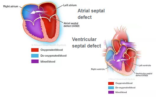
Normally, the left side of the heart only pumps blood to the body, and the heart’s right side only pumps blood to the lungs.  However, sometimes you can be born with a hole or defect in the muscular wall between the right and left sides of the heart (septum) which allows the blood to cross between the right and left chambers of the heart.  An atrial septal defect (ASD) is a condition where the muscular wall between the heart’s two upper chambers of the heart (septum) is affected; where a ventricular septal defect (VSD) affects the muscular wall between the two lower chambers of the heart.  Sometimes open heart surgery is required to close these holes either by placing stitches or with a patch.

Patent Ductus Arteriosus

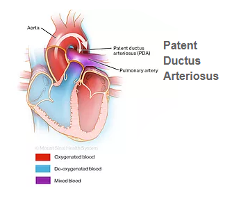
Patent ductus arteriosus is when there is an abnormal connection between the aorta and the pulmonary artery that is present at birth. While in the womb, the aorta and the pulmonary artery in the foetus are connected by a vessel, the ductus arteriosus, as a part of foetal circulation which seals off after birth. If the vessels fail to close after birth it leads to the condition patent ductus arteriosus which causes mixing of blood in the aorta and the pulmonary artery.   Surgery involves making a small surgical cut on the left side of the chest. The surgeon finds the Patent ductus arteriosus vessel, and then ties it off (ligation) or clips it.

Coarctation of the Aorta

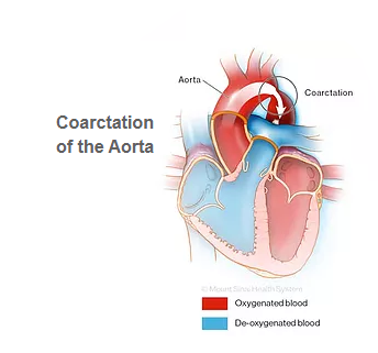
Coarctation of the aorta is a common congenital heart defect where a part of the aorta has a very narrow section. This results in restricted blood flow through the aorta. Symptoms can include shortness of breath, headache, chest pain, exercise intolerance, bleeding from the nose, and leg pain.  To repair this, the surgeon will cut into the wall of the narrow section and make it bigger with a patch.

Pulmonary Artery Stenosis

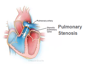
Pulmonary artery stenosis is a congenital heart defect caused by the narrowing of the pulmonary artery that carries impure blood into the lungs. This makes the flow of blood to the lungs difficult.

Tetralogy of Fallot

Tetralogy of Fallot is a rare, congenital heart defect which is a group of four heart defects including large ventricular septal defect, pulmonary stenosis, right ventricular hypertrophy, and an overriding aorta. In Tetralogy of Fallot, the aorta is located between the left and right ventricles, above the ventricular septal defect.​

The surgery involves closing the ventricular septal defect with a patch; opening the pulmonary valve and removing the thickened muscle (stenosis); and placing a patch on the right ventricle and main pulmonary artery to improve blood flow to the lungs.